Discover the causes, symptoms and treatment options for tendonitis to better understand and prevent this inflammation of the tendons.
What is tendonitis?
Tendonitis is a painful inflammation of a tendon, often related to overuse, trauma, or lifestyle factors such as poor diet and physical activity. It is characterised by an inflammatory response initiated by mechanical stress factors and pro-inflammatory signals. This inflammation leads to pain, impaired tendon function and, in some cases, calcification(1).
What are the different types of tendonitis?
There are several types of tendonitis, each occurring in specific areas of the body and caused by different factors. Here are some common types of tendonitis:
Mild and acute tendonitis:
Tendonitis caused by overload mainly affects the rotator cuff, the medial and lateral epicondyles of the elbow, the patellar tendon and the Achilles tendon. In most cases, they can be treated effectively with rest, cessation of activities involving the tendon, and simple measures such as ice application and eccentric strengthening exercises, especially if diagnosed at an early stage(16).
Chronic tendonitis:
Chronic or poorly treated tendonitis is associated with tendon degeneration, characterised by increased turnover and remodelling of its extracellular matrix. This makes it difficult to heal without appropriate treatment. For example, studies have shown that chronic Achilles tendonitis is often resistant to traditional treatments, and to promote adequate healing, a rehabilitation programme should include specific eccentric strengthening exercises(17).
Which parts of the body are most often affected?
Shoulder tendonitis (rotator cuff)
Tendonitis of the rotator cuff, a set of 4 tendons in the shoulder, is often caused by repetitive movements or trauma. This region is particularly affected by calcific tendonitis, characterised by calcium deposits in the tendons. These deposits cause a major inflammatory reaction, leading to intense, acute pain, which is often present at the onset of the disease. However, the deposits may be asymptomatic in 20% of cases(2,23).
Tendonitis of the wrist
De Quervain's tenosynovitis is an inflammation that affects the tendons located at the radial edge of the wrist, more specifically the tendons of the long abductor muscle and the short extensor muscle of the thumb. This condition is characterised by the formation of fibrous tissue that exerts pressure and compression on the tendons, which can lead to pain and difficulty in moving the thumb. It is frequently seen in adults of working age (prevalence 1.3% in women and 0.5% in men), with a peak in frequency between the ages of 40 and 60. Possible causes include acute injury, repetitive and forced movements of the wrist and thumb resulting in increased frictional forces or microtrauma, inflammatory diseases or anatomical variations(24).
Knee tendonitis
Patellar tendinopathy, commonly known as ‘jumper's knee’, is a degenerative disorder rather than an inflammatory disease, making the term ‘tendonitis’ inappropriate. This condition mainly affects athletes who play sports requiring frequent jumping, such as volleyball and basketball, generating a considerable load of energy in the extensor mechanism. It affects around 8.5% of athletes, with a much higher prevalence in men (10.2%) than in women (6.4%). The main risk factors associated with patellar tendinopathy include younger age, greater body stature and higher body weight. Non-surgical treatment of this condition focuses on eccentric exercises, and in cases refractory to non-surgical treatment, surgical intervention is indicated (25,26).
Elbow tendonitis
Lateral epicondylitis, often called ‘tennis elbow’, is a condition characterised by pain and tenderness in the elbow, due to damage to the tendons of the forearm muscles that attach to the lateral epicondyle of the elbow. It affects between 1% and 3% of the adult population, mainly middle-aged people, with no gender predominance. It is frequently caused by activities requiring excessive and repetitive use of the forearm muscles, such as tennis (up to 50% of cases), playing a musical instrument or manual work. It can also result from an acute injury, such as a direct trauma to the lateral epicondyle. Non-surgical treatments, which are effective in 90% of cases, include rest, physiotherapy, orthoses, non-steroidal anti-inflammatory drugs and corticosteroid injections. Surgery is generally considered for patients whose symptoms persist despite well-managed non-surgical treatments(28,29).
1/4
What causes tendonitis?
Modifiable factors
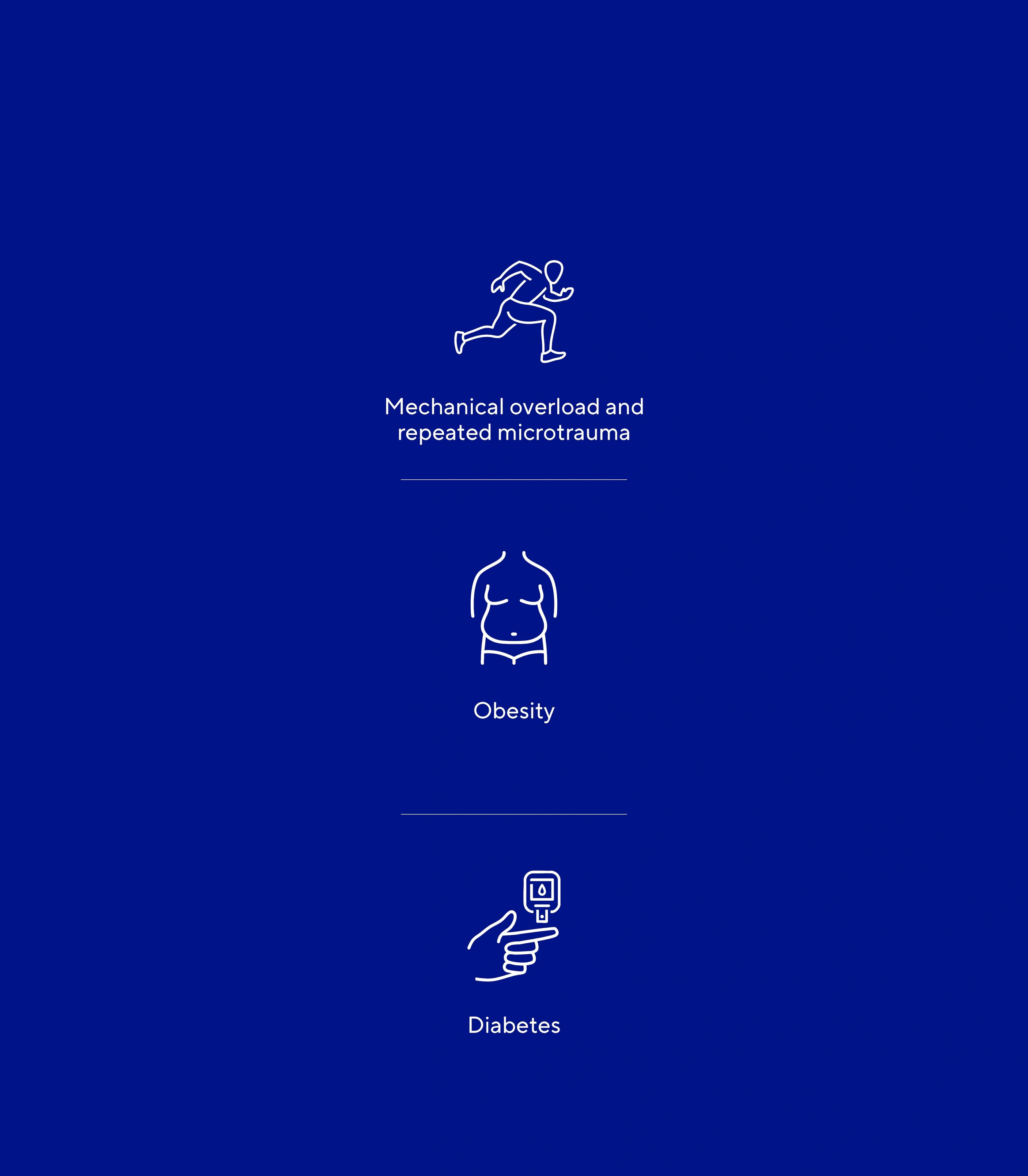
Mechanical overload and repeated microtrauma One of the main causes of tendonitis is the repetition of mechanical movements, particularly in the context of sporting or professional activities. These movements put excessive strain on the tendons, leading to inflammation and pain. Repeated microtrauma can also gradually damage tendon tissue, leading to chronic inflammation, as is frequently seen in athletes(5).
Obesity Obesity has been shown to have deleterious effects on tendons, with a multifactorial aetiology including mechanical overload due to increased body weight and an increase in systemic bioactive peptides, such as leptin, which contribute to a chronic low-grade inflammatory state(6).
Diabetes Studies show that tendinopathies are more frequent in diabetics than in non-diabetics. Diabetes promotes structural changes in tendons, such as a statistically significant increase in their size and irregularities in their fibre bundles(6).
Non-modifiable factors
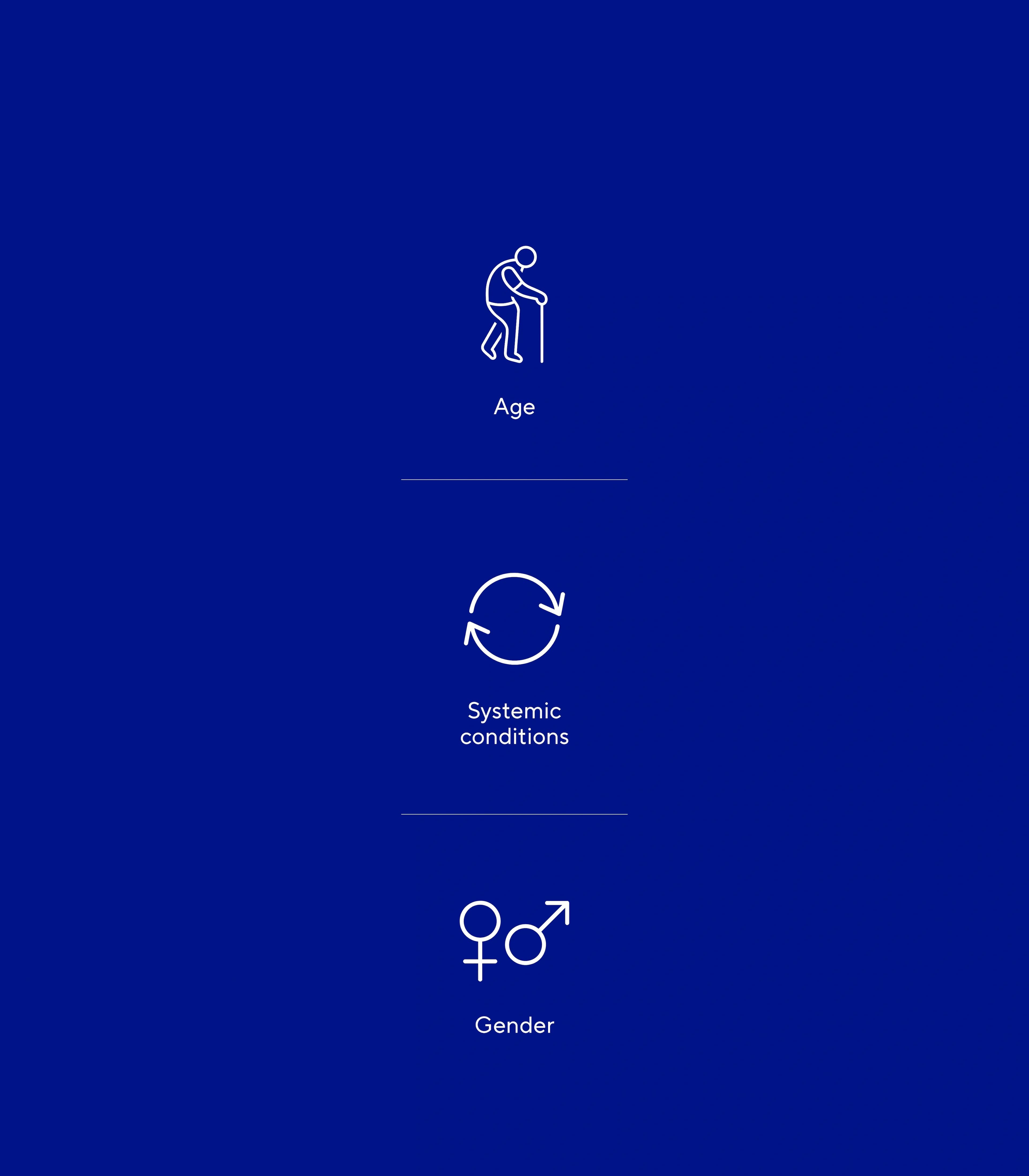
Age Ageing is a major risk factor because tendon vascularisation decreases with age, slowing down their healing. This reduction in blood supply limits the inflammation required for repair, thereby impairing tendon healing and making them more vulnerable to degeneration. In addition, with age, tendons lose their elasticity, making them more vulnerable to injury(6).
Systemic conditions Certain inflammatory diseases, such as lupus, increase the risk of developing tendonitis. These conditions cause chronic inflammation that affects the tendons(7).
Gender Studies show that in physically inactive adults, men have tendons that are subjected to greater stress, making them more likely to develop Achilles tendonitis due to the morphological properties of the tendon(8).
What are the symptoms and consequences?
Common symptoms of tendonitis:
Pain Pain is the main symptom of tendonitis, often localised to the affected tendon. For example, in calcific tendonitis, the pain can be acute and intense, especially when the calcium deposit becomes inflamed(9).
Swelling and inflammation Tendonitis is a condition that often causes swelling around muscles and bones. It most often affects the shoulder, elbow, wrist, hip, knee or ankle(10).
Joint stiffness Repetition of the same movements and excessive strain on the joints, particularly in the elderly, increases the risk of tendonitis. This phenomenon, which is common among manual workers (carpenters, gardeners) and high-level sportsmen and women, is characterised by stiffness and reduced mobility in the affected area(11).
Consequences of tendonitis:
Impairment of function By causing pain and inflammation in the tendon, tendonitis frequently leads to functional limitations in the joint concerned, manifested by a reduction in range of movement and mobility. For example, shoulder tendonitis can restrict elevation of the upper limb(12).
A risk of tendon rupture In the most serious cases of tendonitis, especially when poorly treated, the tendon can rupture spontaneously, particularly in the Achilles tendon. This rupture, often linked to degeneration of the tendon tissue, generally requires surgery(13).
In conclusion, tendonitis leads to pain and loss of function, and can even lead to rupture of the tendon in the event of complications.
FAQ about tendonitis
Our medical team answers the questions you may have.
It is possible for some tendonitis to resolve itself, but this depends on a number of factors, including the severity of the tendonitis, its location and the patient's physical activity. In fact, in many cases, tendonitis, especially chronic tendonitis, does not completely heal without therapeutic intervention.
The length of time it takes for tendonitis to heal varies and depends on a number of factors, such as the anatomical location, the severity of the injury and the treatment methods used. Typical healing times are as follows:
Mild tendonitis: Acute tendonitis, if treated promptly with rest and appropriate care, can resolve in a few weeks, generally between 2 and 6 weeks(18).
Chronic tendonitis: Chronic forms of tendonitis, which last for more than three months, can take much longer to heal. For example, studies show that chronic elbow tendonitis generally takes between 3 and 6 months to heal, after a comprehensive care programme including patient education has been put in place(18).
Calcific tendonitis: In the case of calcific tendonitis, the duration of the pathology can vary considerably, ranging from a few months to several years. This depends largely on the resorption of the calcification, a process that is difficult to predict. Studies have shown that non-surgical treatments, such as low-energy shockwave therapy, can accelerate healing in patients suffering from calcific shoulder tendonitis. Significant improvements were observed in many patients within the first 6 to 12 weeks of this treatment(19).
There are several approaches to treating tendonitis:
Rest and modification of activities to avoid overstressing the tendon(14).
Wearing an orthosis to relieve the affected area(15).
Re-education exercises (eccentric) to promote healing under supervision(14).
Anti-inflammatory drugs (NSAIDs) to reduce short-term pain(14).
Platelet-rich plasma (PRP) injections to improve recovery in some cases(14,15).
Ultrasound therapy to stimulate tissue repair(14).
Surgery as a last resort if conservative treatments fail(16).
Mueller, A., Brockmueller, A., Kunnumakkara, A., & Shakibaei, M. (2022). Modulation of Inflammation by Plant-Derived Nutraceuticals in Tendinitis. Nutrients, 14. https://doi.org/10.3390/nu14102030.
Elshewy, M. (2016). Calcific tendinitis of the rotator cuff.. World journal of orthopedics, 7 1, 55-60 . https://doi.org/10.5312/wjo.v7.i1.55
Choi, H., Choi, S., Kim, J., Song, M., Shim, K., Lim, Y., Kim, H., Park, K., & Kim, S. (2020). Enhanced tendon restoration effects of anti-inflammatory, lactoferrin-immobilized, heparin-polymeric nanoparticles in an Achilles tendinitis rat model.. Carbohydrate polymers, 241, 116284 . https://doi.org/10.1016/j.carbpol.2020.116284.
Merolla, G., Dave, A., Paladini, P., Campi, F., & Porcellini, G. (2014). Ossifying tendinitis of the rotator cuff after arthroscopic excision of calcium deposits: report of two cases and literature review. Journal of Orthopaedics and Traumatology : Official Journal of the Italian Society of Orthopaedics and Traumatology, 16, 67 - 73. https://doi.org/10.1007/s10195-014-0309-8
Kannus, P. (1997). Etiology and pathophysiology of chronic tendon disorders in sports. Scandinavian Journal of Medicine & Science in Sports, 7. https://doi.org/10.1111/j.1600-0838.1997.tb00123.x.
Andrew E. Federer, John R. Steele, Travis J. Dekker, Jordan L. Liles, Samuel B. Adams, Tendonitis and Tendinopathy: What Are They and How Do They Evolve?, Foot and Ankle Clinics, Volume 22, Issue 4, 2017, Pages 665-676, ISSN 1083-7515, ISBN 9780323552769, https://doi.org/10.1016/j.fcl.2017.07.002(https://www.sciencedirect.com/science/article/pii/S1083751517300724)
Jarrot, P., Leone, J., Brochot, P., & Pennaforte, J. (2015). Achilles tendinitis in systemic lupus erythematosus: search for an associated inflammatory disease. Lupus, 24, 1318 - 1320. https://doi.org/10.1177/0961203315576590.
Zhang, X., Deng, L., Xiao, S., Li, L., & Fu, W. (2021). Sex Differences in the Morphological and Mechanical Properties of the Achilles Tendon. International Journal of Environmental Research and Public Health, 18. https://doi.org/10.3390/ijerph18178974.
Williams, A., Stang, T., Fritz, J., & Papp, D. (2016). Calcific Tendinitis of the Gluteus Maximus in a Golfer.. Orthopedics, 39 5, e997-e1000 . https://doi.org/10.3928/01477447-20160616-08.
Lofthouse, R. (1957). Bursitis and tendinitis.. Canadian services medical journal, 13 3, 155-9 .
Zibis, A., Giannis, D., Malizos, K., Kitsioulis, P., & Arvanitis, D. (2013). Acute calcific tendinitis of the longus colli muscle: case report and review of the literature. European Spine Journal, 22, 434-438. https://doi.org/10.1007/s00586-012-2584-5.
Manifestation of Autoimmune Hepatitis. ARC Journal of Orthopedics. https://doi.org/10.20431/2456-0588.0402004.)
Józsa, L., Réffy, A., Kannus, P., Demel, S., & Elek, E. (2004). Pathological alterations in human tendons. Archives of Orthopaedic and Trauma Surgery, 110, 15-21. https://doi.org/10.1007/BF00431359.
Kane, S. F., Olewinski, L. H., & Tamminga, K. S. (2019, août 1). Management of Chronic Tendon Injuries. AAFP. https://www.aafp.org/pubs/afp/issues/2019/0801/p147.html
Kim, Y., Lee, H., Kim, Y., & Kong, C. (2014). Which method is more effective in treatment of calcific tendinitis in the shoulder? Prospective randomized comparison between ultrasound-guided needling and extracorporeal shock wave therapy.. Journal of shoulder and elbow surgery, 23 11, 1640-6 . https://doi.org/10.1016/j.jse.2014.06.036.
Wilson, J., & Best, T. (2005). Common overuse tendon problems: A review and recommendations for treatment.. American family physician, 72 5, 811-8 .
Stanish, W., Rubinovich, R., & Curwin, S. (1986). Eccentric Exercise in Chronic Tendinitis. Clinical Orthopaedics and Related Research, 45, 65–68. https://doi.org/10.1097/00003086-198607000-00014.
Gabel, G. (1999). Acute and chronic tendinopathies at the elbow.. Current opinion in rheumatology, 11 2, 138-43 . https://doi.org/10.1097/00001433-200002000-00010.
Wang, C., Ko, J., & Chen, H. (2001). Treatment of Calcifying Tendinitis of the Shoulder With Shock Wave Therapy. Clinical Orthopaedics and Related Research, 1, 83-89. https://doi.org/10.1097/00003086-200106000-00011.
Loew, L., Brosseau, L., Tugwell, P., Wells, G., Welch, V., Shea, B., Poitras, S., Angelis, G., & Rahman, P. (2014). Deep transverse friction massage for treating lateral elbow or lateral knee tendinitis.. The Cochrane database of systematic reviews, 11, CD003528 . https://doi.org/10.1002/14651858.CD003528.pub2.
Stefansson, S., Brandsson, S., Langberg, H., & Arnason, A. (2019). Using Pressure Massage for Achilles Tendinopathy: A Single-Blind, Randomized Controlled Trial Comparing a Novel Treatment Versus an Eccentric Exercise Protocol. Orthopaedic Journal of Sports Medicine, 7. https://doi.org/10.1177/2325967119834284.
Chaves, P., Simões, D., Paço, M., Silva, S., Pinho, F., Duarte, J., & Ribeiro, F. (2020). Deep Friction Massage in the Management of Patellar Tendinopathy in Athletes: Short-Term Clinical Outcomes.. Journal of sport rehabilitation, 1-6 . https://doi.org/10.1123/jsr.2019-0046.
Merolla, G., Singh, S., Paladini, P. et al. Calcific tendinitis of the rotator cuff: state of the art in diagnosis and treatment. J Orthopaed Traumatol 17, 7–14 (2016). https://doi.org/10.1007/s10195-015-0367-6
The side of my wrist hurts. (n.d.). Australian Journal of General Practice. https://www1.racgp.org.au/ajgp/2019/november/side-of-my-wrist-hurts
Figueroa, D., Figueroa, F., & Calvo, R. (2016). Patellar Tendinopathy: Diagnosis and Treatment. Journal of the American Academy of Orthopaedic Surgeons, 24, e184–e192. https://doi.org/10.5435/JAAOS-D-15-00703.
Zwerver J, Bredeweg SW, van den Akker-Scheek I. Prevalence of Jumper’s Knee Among Nonelite Athletes From Different Sports: A Cross-Sectional Survey. The American Journal of Sports Medicine. 2011;39(9):1984-1988. doi:10.1177/0363546511413370
Nichols, A. (1989). Achilles Tendinitis In Running Athletes. The Journal of the American Board of Family Medicine, 2, 196 - 203. https://doi.org/10.3122/jabfm.2.3.196.
Vaquero-Picado, A., Barco, R., & Antuña, S.A. (2016). Lateral epicondylitis of the elbow. EFORT Open Reviews, 1, 391 - 397.
Cutts, S., Gangoo, S., Modi, N., & Pasapula, C. (2020). Tennis elbow: A clinical review article.. Journal of orthopaedics, 17, 203-207 . https://doi.org/10.1016/J.JOR.2019.08.005.